
Tuesday, April 26, 2011

Monday, April 18, 2011
Current Debates in Animal Evolution: Lecture 1 - Why Evolution (Part 1)
I'm currently teaching a seminar at UCLA for the Spring quarter. My idea was to create a course that focuses on current scientific research in animal evolution. The first part of my first lecture is
on youtube; you can check it out below:
on youtube; you can check it out below:
Friday, April 8, 2011
Self-Assembled Eyes Made Simple
 Yokishi Sasai and his collaborators have been studying how mouse embryonic stem cells can be encouraged to develop into organs, particularly eyes. In a recent issue of the journal Nature, Eiraku et al. describe a way to culture these stem cells in a new medium, which allows the cells to develop into dynamic, three-dimensional shapes.
When Eiraku et al. encouraged the stem cells to express an eye-inducing gene (retinal homeobox, also called Rx), a surprising result occurred. Little vesicles developed from the aggregate of stem cells, and each vesicle self-organized into a simple optic cup, which is the first step towards developing a complex eye in mice (and ourselves):

The reason this result is so surprising is that many scientists have thought that patterning a structure as complicated as the optic cup would require chemical cues from other parts of the developing head, or from the eye lens that normally develops in alongside the optic cup.
In this next image, you can see how the developing optic cup begins expressing different genes in different cells, Rx is expressed in the cells facing outward, while another well-known eye-inducing gene, Pax6, begins to be expressed in the cells towards the back:

The changes in gene expression correlate with changes in cell flexibility. Cells towards the back (seen above in red) become more ridged, while cells facing outwards (in green) become more flexible. This allows the cells facing outwards to buckle in, creating a self-organized cup.
There are some interesting evolutionary consequences to this study. Some of the simplest animal eyes, such as the ones I study in the jellyfish Aurelia, are simple cup eyes that are structurally similar to the optic cups described above:

Aurelia does not have a head or a lens to induce development of the optic cup, so it is interesting to see that mouse stem cells can self-assemble optic cups without the help of other body parts.
Do mouse optic cups develop in the same manner as jellyfish? We do not know yet if Aurelia has the gene Rx, but a relative of Aurelia—the sea anemone Nematostella—definitely does (Mazza et al. 2010). Our lab also has evidence that the optic cups of Aurelia do express a gene similar to Pax6 as well. Whether the complex eyes of mice are modifications of the simple cup eyes of animals like jellyfish is going to require more research, but it is an intriguing possibility.
Works Cited
Mazza ME, Pang K, Reitzel AM, Martindale MQ, & Finnerty JR (2010). A conserved cluster of three PRD-class homeobox genes (homeobrain, rx and orthopedia) in the Cnidaria and Protostomia. EvoDevo, 1 (1) PMID: 20849646
Mototsugu Eiraku, Nozomu Takata, Hiroki Ishibashi, Masako Kawada, Eriko Sakakura, Satoru Okuda, Kiyotoshi Sekiguchi, Taiji Adachi, Yoshiki Sasai. Self-organizing optic-cup morphogenesis in three-dimensional culture. Nature, 2011; 472 (7341): 51 DOI: 10.1038/nature09941
Saturday, March 19, 2011
Of Chicken Wings and Dinosaur Hands
The idea that birds evolved from dinosaurs dates back to the 1800’s (Cope 1867; Huxley 1870), but it was largely abandoned for almost a century. The bird-dinosaur connection didn’t start gaining traction again until the 1960’s and 70’s, largely through John Ostrom’s discovery and detailed studies of particularly avian dinosaurs such as Deinonychus (a cousin of the more famous Velociraptor, see Ostrom 1976). The hypothesis was heavily vindicated in the mid-2000’s with the discovery of a variety of dinosaurs (largely from China) sporting feathers and/or protofeathers. It’s hard to see a fossil like the incredibly well-preserved specimen below, and not be struck by the clear connection between dinosaurs and birds:
Today, it is generally considered common knowledge that birds evolved from dinosaurs (in fact, a recent commenter on one of my YouTube videos chastised me for implicitly suggesting otherwise). However, there has been one major sticking point in this hypothesis. Bird wings and dinosaur hands both show a reduction in the number of fingers, going from an ancestral five-fingered hand to a three-fingered hand. But while paleontological evidence suggests that the bird-like dinosaurs (like Deinonychus) lost digits five and four, embryological evidence suggests that modern birds lost digits one and five:

This discrepancy has led some scientists to challenge the whole idea that birds evolved from advanced dinosaurs (e.g. Feduccia et al. 2005). But in a recent issue of the journal Science, Tamura et al. revisited the problem, and found new evidence that the bird hand actually retains digits one, two, and three, just like dinosaurs.
In four-legged vertebrates, digits (a.k.a. fingers and toes) begin to develop when the limb is little more than a bud sticking out of the embryo (you can see some images of developing limb buds in my post How to Build a Marsupial). Typically, the first visible digit in the developing limb bud is digit four. Scientists have traditionally argued that the first visible digit in chickens is digit four, as it is in reptiles and mammals, meaning the three remaining fingers in bird wings would be digits two, three, and four.
Tamura et al. challenged this hypothesis using detailed cell-labeling and tissue graft experiments to see how gene expression controls digit specification. In most limb buds, digits four and five develop in a region of cells collectively called the zone of polarizing activity (ZPA). These cells release a gene called sonic hedgehog, which forms a gradient that moves up the limb bud, specifying the formation of digits three and two. Below is a figure that sums up the results of the research performed by Tamura et al. It compares development of a mouse limb, a chicken leg (hindlimb) and a chicken wing (forelimb):

Don’t be overwhelmed by the amount of data in this image; start by noticing the difference between the theoretical position (P) of digits shared in all early limb buds, and the actual digit (D) that develops. In the chicken leg (hindlimb), position four (P4) stays within the ZPA (colored light blue) so it takes on the identity of the fourth digit (D4), just like in the mouse. But in the chicken wing (forlimb), position four (P4) moves out of the ZPA, so it takes on the identity of the third digit (D3). Similarly, because the ZPA has shifted, the gradient of sonic hedgehog (the grey curve that says “SHH”) now travels to position four and position three, turning them into digits two and three.
This work shows that the presence of the first visible digit is not a reliable way of determining what the identity of each digit actually is. In a sense, this might seem like semantics, but by showing that chicken fingers have the same identity as dinosaur fingers refutes that last major challenge against the bird-dinosaur debate. Gives you something to think about next time you enjoy some buffalo wings…
Works Cited
Cope E. D. (1867). Account of extinct reptiles which approach birds. Proceedings of the Academy of Natural Sciences of Philadelphia: 234-235.
Feduccia, A., Lingham-Soliar, T., & Hinchliffe, J. (2005). Do feathered dinosaurs exist? Testing the hypothesis on neontological and paleontological evidence Journal of Morphology, 266 (2), 125-166 DOI: 10.1002/jmor.10382
Huxley, T. (1870). Further Evidence of the Affinity between the Dinosaurian Reptiles and Birds Quarterly Journal of the Geological Society, 26 (1-2), 12-31 DOI: 10.1144/GSL.JGS.1870.026.01-02.08
Ostrom, J. (1976). Archaeopteryx and the origin of birds Biological Journal of the Linnean Society, 8 (2), 91-182 DOI: 10.1111/j.1095-8312.1976.tb00244.x
Tamura K, Nomura N, Seki R, Yonei-Tamura S, & Yokoyama H (2011). Embryological evidence identifies wing digits in birds as digits 1, 2, and 3. Science (New York, N.Y.), 331 (6018), 753-7 PMID: 21311019
Tuesday, March 8, 2011
How to Build an Arthropod (An Arthropod Leg, at Least)
Liu J, Steiner M, Dunlop JA, Keupp H, Shu D, Ou Q, Han J, Zhang Z, & Zhang X. (2011) An armoured Cambrian lobopodian from China with arthropod-like appendages. Nature, 470(7335), 526-30.


Liu J, Steiner M, Dunlop JA, Keupp H, Shu D, Ou Q, Han J, Zhang Z, & Zhang X (2011). An armoured Cambrian lobopodian from China with arthropod-like appendages. Nature, 470 (7335), 526-30 PMID: 21350485
Pechenik, J. A. (2010). Biology of the Invertebrates (Sixth Edition). New York: McGraw-Hill Higher Education.

If you’ve ever wondered what a “typical” animal looks like, here it is:
Beetles are the largest group of animals in the world, constituting 400,000 species, or about 40% of all known animals on earth. Beetles and other insects are part of the phylum Arthropoda, which encompasses animals that have an exoskeleton and jointed appendages. Besides the insects, crabs, spiders, centipedes, scorpions, and the extinct trilobites are also arthropods. Together, arthropods account for 80% of all known animals.
Genetic evidence suggests that the closest living relatives of arthropods are the Onycophorans, or velvet worms. These animals show segmentation like the arthropods, but they lack rigid bodies or limbs. Interestingly, the specialized, jointed appendages of arthropods seems to have been critical to their evolutionary success; although over a million species of arthropods are known, only about 70 living species of onycohporans have been described (Pechenik 2010).

Recently, a team of paleontologists from China and Germany published some exceptionally preserved Cambrian fossils that show a possible link between the onycohporan and arthropod bodyplans. The animal, called Diania cactiformis, has a soft body like an onycohporan, but hardened, jointed limbs like arthropods. Below is an image of one of the fossils from the publication, as well as a reconstruction of what the animal may have looked like:
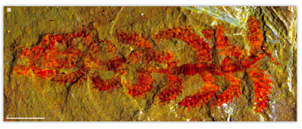

Diania is not the first fossil animal to bridge the gap between onycohporans and arthropods. During the Cambrian, a number of animals generically lumped together as lobopods evolved, which had soft, worm-like bodies with varying degrees of armor on their shoulders and legs (including Hallucigenia, which wins my nomination for greatest animal name in history). But Diania shows more characteristics in common with true arthropods than any previously described lobopod.
The authors of this paper are hesitant to say that this conclusively shows that hardened arthropod limbs evolved before hardened arthropod bodies, or speculate on why complex, hardened limbs might have evolved first. Whatever the case, Diania is an incredible example that “missing links” in the fossil record do not typically look like a perfectly intermediate blend of two animals. Instead they often have an unexpected mosaic of features, and show unique adaptations suited to their distinct environments.
Works Cited
Pechenik, J. A. (2010). Biology of the Invertebrates (Sixth Edition). New York: McGraw-Hill Higher Education.
Thursday, February 17, 2011
A Bio-Blueprints Primer: Paraphyly and Last Common Ancestors
There are a couple of papers that I want to discuss soon that involve the topic of paraphyly. Instead of trying to cram background and a paper review into one post, I am trying a new method, where I present a quick primer of a concept that can be referred back to in later posts. Perhaps that will make this post none too interesting, but I hope it will be useful in the future.
It’s hard not to call the jellyfish that I study “primitive”. Certainly jellyfish are much simpler than most animals; they lack organs, a central nervous system, or a middle layer of cells. Living jellyfish also look similar to ones in the fossil record, so it is probably fair to say they haven’t changed much during the course of evolution. However, the reason I try to stop myself from calling them “primitive” is that it gives the false impression that our ancestors looked like a jellyfish.
For example, let’s take a simple evolutionary tree (or phylogeny), showing the relationship between a jellyfish and a human, and ask ourselves what the common ancestor of these two animals looked like:

Given the information I presented about jellyfish, you might think that the last common ancestor looked like the simple jellyfish. However, both the jellyfish and the human have been evolving for an equal amount of time, so the last common ancestor of the two animals might look nothing like either species:

But let’s say we add a second species of jellyfish to our phylogeny, and we get this result:

In this scenario, some jellyfish are more closely related to humans than they are to other jellyfish. This means the last common ancestor of the creatures we call “jellyfish” would also include the ancestor of human beings. If this tree were correct, scientists would say that “jellyfish” are a paraphyletic group, meaning that you cannot capture the last common ancestor of jellyfish without also including other animals that we would not normally call jellyfish.
The nice thing about paraphyletic groups is that they give you a much better idea of what the last common ancestor looked like. In my jellyfish example, the last common ancestor of these three animals either looked like a jellyfish, or both “jellyfish” lineages independently evolved all of the characteristics that jellyfish have in common (which is unlikely).
There is no scientific evidence that the phylogeny I presented is true. It was just an example of the principle. However, one example of a paraphyletic group that scientists do think is real is the “reptiles”:

For a long time, some scientists have argued that crocodiles are more closely related to birds then they are to other reptiles (lizards, snakes, or turtles). This is because fossil evidence suggests that crocodiles are evolutionary cousins of the dinosaurs, and birds evolved from dinosaurs. DNA evidence supports this hypothesis, meaning that the term “reptile” is paraphyletic.
This phylogeny provides good evidence that the last ancestor of birds and crocodiles probably was cold-blooded, and had four legs, scaly skin, and a tail. Of course, if we only looked at living animals, we would have no idea of the bizarre creatures that connect the two lineages, which is why paleontology is so important:

In coming posts, I will discuss the growing body of genetic evidence that suggests that many groups of animals are paraphyletic, and how this provides a better understanding of the direction of evolutionary change.
Monday, February 14, 2011
Subscribe to:
Posts (Atom)