Tuesday, July 26, 2011

4th of July Festival

Because Woods Hole, MA is home to both an Oceanographic Institute and the Marine Biological Laboratory, most of the people in this small town have some connection to the scientific community. As a result, the fourth of July festival in Woods Hole, MA is a celebration of uninhibited science-geekery.

Some of the highlights included the neurobiology course:


Microbial diversity, with their giant squid:


And my personal favorite, Mendel with his peas:


Bringing up the rear was our own Embryology course.  We have a time-honored tradition of performing gastrulation through interpretive dance, which is quite possibly the best way to show gastrulation:


If that wasn’t self explanatory, let me explain.

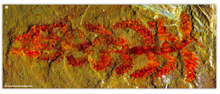
If you’re not familiar with gastrulating, it’s a process during development where animals generate their gut.  All animals start out as a single cell, the fertilized egg.  This cell divides over and over again until it forms a hollow sphere of cells, called a blastula.  Some cells from the blastula then move inside the hollow sphere, creating a sphere with multiple layers of cells.  The inside layer, called the endoderm, will form the gut and some organs.  The middle layer, or mesoderm, will form the musculature and bones.  The outer layer, or ectoderm, forms skin and the nervous system.

This image shows endoderm (in red) moving into the blastula.  The ectoderm is in orange, the mesoderm is not shown.  Image taken from Wikipedia

The three different colors of our shirts represent the three germ layers: blue for ectoderm, red for mesoderm, yellow for endoderm.  In this particular display, we are performing gastrulation as it occurs in the sea urchin (that was obvious, right?) We started out as a blastula (a hollow ball of cells), and then invaginated to create the three layers of tissue. Finally, some of the mesoderm cells start to form spicules, which create the skeleton of the urchin.

Sea urchins were only the beginning. We also performed gastrulation as it occurs in frogs, nematode worms, fruit flies, as well as chaotic cleavage (like you find in some sea anemones and jellyfish) and chicken neurulation for good measure.  The parade lasted less than an hour, and only traveled a few blocks, but we got as many gastrulations as we could in there. 

If you’re ever in the area during the fourth of July, I highly recommend you check out the Woods Hole parade.  Their were a lot of people, and a lot of energy (including an epic water gun fight).  


Tuesday, July 5, 2011

The Course T-shirts have been Finalized

And were designed by yours truly.  Unfortunately, the quality of the images went down during processing; I'll try to get some higher resolution images up soon. Take a look:

Here's the front...

...and the back

Tuesday, June 28, 2011

Greetings From Woods Hole, Massachusetts


Sorry for the long delay in posting.  I am currently taking the Embryology course at the Marine Biological Laboratory. Actually, I’ve been here since the beginning of June, but the work schedule has made it difficult to get any of these blog posts up.  Every day we begin with lectures around 9am, and we often do laboratory work well past 1 or 2 in the morning!

However, I have been asked to share my experiences with The Node, a blog run by The Company of Biologists, which runs a number of important journals, including Development and The Journal of Experimental Biology. It’s a great incentive to get these posts out, despite the lack of free time.  But we’re half way done with the course, so I’ll have a lot of catching up to do. 

On June 7th, Nicole King from Berkeley came to teach us how to work with choanoflagellates and sponges.  I have gotten to spend time with Nicole previously because we work on the same NASA astrobiology grant (which you can find more about here or here) , but this was my first chance to get some hands-on work with her model systems. 

Nicole focuses on sponges and choanoflagellates to learn more about the early evolution of animals.  Choanoflagellates are not actually animals, but DNA evidence suggests that they are the animals’ closest living relatives (e.g. King et al 2008).  An individual choanoflegellate is made up of a single cell. It has a long flagellum, which it uses to swim through the water and to trap bacteria (which it eats) in a collar made up of microvilli.  Below is a diagram of a choanoflagellate, courtesy of ChoanoWiki:


The reason that choanoflagellates have received a lot of attention recently is that they don’t always stay as single cells.  Sometimes, as a choaflagellate divides, the cells stay connected to each other.  The creatures will form a variety of shapes, including long chains and rosettes:

 Image taken from ChoanoWiki.

The fact that these creatures are (1) closely related to the animals, (2) can live as single cells or in mutlicellular groups, and (3) look suspiciously similar to the cells which line the inside of sponges, means that they might provide real insight into the origins of animals.  To find out more about what choanoflagellates can teach us regarding animal evolution, I recommend you look at the papers cited in ChoanoWiki. But for now, I’ll leave you with a very cool picture made by fellow classmate Valerie Virta using antibody staining techniques Nicole taught us.  This is a colony  of choanoflagellates, the blue is staining the bodies of the choanoflagellate bodies, the red is staining the microvillar collar, and the green is staining flagellum (for you technical folks, that’s DAPI, actin, and tubulin):


You can also see a cool (if not particularly informative) I took of a sponge below.  Most of the color is generated from natural florescence coming from the skeleton of the sponge (called spicules).  But you can see the little blue dots, which are the nuclei of cells stained with DAPI. 


Wednesday, May 18, 2011

Evolution of Constraint - What Causes and Breaks Dollo's Law?

ResearchBlogging.org
When something is lost in evolution, it is rarely gained again.  The trend of was first noted in 1893 by the paleontologist Luis Dollo.  One famous example of this trend, often called Dollo’s law, is the number of digits on the hands and feet of vertebrates.  Some of the earliest fossil amphibians have eight or more toes, but that number was quickly reduced to five:


For the last 300 million years, all animals, from humans to whales, dinosaurs to birds, have had five digits or less on their hands and feet. But a good rule of thumb is that any law in biology is going to have exceptions.  Anyone who knows enough about domestic dogs and cats knows that some breeds are found with multiple toes. Here is the paw of a Norwegian Lundehund, six toes and all:


What explains this violation of Dollo’s law? What explains Dollo’s law in the first place?  For the next few posts, I’m going to look at the science behind evolutionary constraint, what causes it, and what happens when it’s broken.


Works Cited


Boisvert CA, Mark-Kurik E, & Ahlberg PE (2008). The pectoral fin of Panderichthys and the origin of digits. Nature, 456 (7222), 636-8 PMID: 18806778


Dollo, L (1893). Les lois de l'évolution. Bull. Soc. Belge Geol. Pal. Hydr, VII:164-166.

Monday, April 18, 2011

Current Debates in Animal Evolution: Lecture 1 - Why Evolution (Part 1)

I'm currently teaching a seminar at UCLA for the Spring quarter.  My idea was to create a course that focuses on current scientific research in animal evolution.  The first part of my first lecture is 
on youtube; you can check it out below:


Friday, April 8, 2011

Self-Assembled Eyes Made Simple


ResearchBlogging.orgYokishi Sasai and his collaborators have been studying how mouse embryonic stem cells can be encouraged to develop into organs, particularly eyes.  In a recent issue of the journal Nature, Eiraku et al. describe a way to culture these stem cells in a new medium, which allows the cells to develop into dynamic, three-dimensional shapes.


When Eiraku et al. encouraged the stem cells to express an eye-inducing gene (retinal homeobox, also called Rx), a surprising result occurred.  Little vesicles developed from the aggregate of stem cells, and each vesicle self-organized into a simple optic cup, which is the first step towards developing a complex eye in mice (and ourselves):


The reason this result is so surprising is that many scientists have thought that patterning a structure as complicated as the optic cup would require chemical cues from other parts of the developing head, or from the eye lens that normally develops in alongside the optic cup.

In this next image, you can see how the developing optic cup begins expressing different genes in different cells, Rx is expressed in the cells facing outward, while another well-known eye-inducing gene, Pax6, begins to be expressed in the cells towards the back:


The changes in gene expression correlate with changes in cell flexibility.  Cells towards the back (seen above in red) become more ridged, while cells facing outwards (in green) become more flexible. This allows the cells facing outwards to buckle in, creating a self-organized cup. 

There are some interesting evolutionary consequences to this study.  Some of the simplest animal eyes, such as the ones I study in the jellyfish Aurelia, are simple cup eyes that are structurally similar to the optic cups described above:


Aurelia does not have a head or a lens to induce development of the optic cup, so it is interesting to see that mouse stem cells can self-assemble optic cups without the help of other body parts. 

Do mouse optic cups develop in the same manner as jellyfish?  We do not know yet if Aurelia has the gene Rx, but a relative of Aurelia­—the sea anemone Nematostella—definitely does (Mazza et al. 2010).  Our lab also has evidence that the optic cups of Aurelia do express a gene similar to Pax6 as well.  Whether the complex eyes of mice are modifications of the simple cup eyes of animals like jellyfish is going to require more research, but it is an intriguing possibility.

Works Cited


Mazza ME, Pang K, Reitzel AM, Martindale MQ, & Finnerty JR (2010). A conserved cluster of three PRD-class homeobox genes (homeobrain, rx and orthopedia) in the Cnidaria and Protostomia. EvoDevo, 1 (1) PMID: 20849646


Mototsugu Eiraku, Nozomu Takata, Hiroki Ishibashi, Masako Kawada, Eriko Sakakura, Satoru Okuda, Kiyotoshi Sekiguchi, Taiji Adachi, Yoshiki Sasai. Self-organizing optic-cup morphogenesis in three-dimensional culture. Nature, 2011; 472 (7341): 51 DOI: 10.1038/nature09941


Saturday, March 19, 2011

Of Chicken Wings and Dinosaur Hands

ResearchBlogging.orgThe idea that birds evolved from dinosaurs dates back to the 1800’s (Cope 1867; Huxley 1870), but it was largely abandoned for almost a century. The bird-dinosaur connection didn’t start gaining traction again until the 1960’s and 70’s, largely through John Ostrom’s discovery and detailed studies of particularly avian dinosaurs such as Deinonychus (a cousin of the more famous Velociraptor, see Ostrom 1976). The hypothesis was heavily vindicated in the mid-2000’s with the discovery of a variety of dinosaurs (largely from China) sporting feathers and/or protofeathers. It’s hard to see a fossil like the incredibly well-preserved specimen below, and not be struck by the clear connection between dinosaurs and birds:
Today, it is generally considered common knowledge that birds evolved from dinosaurs (in fact, a recent commenter on one of my YouTube videos chastised me for implicitly suggesting otherwise). However, there has been one major sticking point in this hypothesis.  Bird wings and dinosaur hands both show a reduction in the number of fingers, going from an ancestral five-fingered hand to a three-fingered hand.  But while paleontological evidence suggests that the bird-like dinosaurs (like Deinonychus) lost digits five and four, embryological evidence suggests that modern birds lost digits one and five:
This discrepancy has led some scientists to challenge the whole idea that birds evolved from advanced dinosaurs (e.g. Feduccia et al. 2005). But in a recent issue of the journal Science, Tamura et al. revisited the problem, and found new evidence that the bird hand actually retains digits one, two, and three, just like dinosaurs.

In four-legged vertebrates, digits (a.k.a. fingers and toes) begin to develop when the limb is little more than a bud sticking out of the embryo (you can see some images of developing limb buds in my post How to Build a Marsupial). Typically, the first visible digit in the developing limb bud is digit four.  Scientists have traditionally argued that the first visible digit in chickens is digit four, as it is in reptiles and mammals, meaning the three remaining fingers in bird wings would be digits two, three, and four.

Tamura et al. challenged this hypothesis using detailed cell-labeling and tissue graft experiments to see how gene expression controls digit specification.  In most limb buds, digits four and five develop in a region of cells collectively called the zone of polarizing activity (ZPA).  These cells release a gene called sonic hedgehog, which forms a gradient that moves up the limb bud, specifying the formation of digits three and two.  Below is a figure that sums up the results of the research performed by Tamura et al.  It compares development of a mouse limb, a chicken leg (hindlimb) and a chicken wing (forelimb):
Don’t be overwhelmed by the amount of data in this image; start by noticing the difference between the theoretical position (P) of digits shared in all early limb buds, and the actual digit (D) that develops.  In the chicken leg (hindlimb), position four (P4) stays within the ZPA (colored light blue) so it takes on the identity of the fourth digit (D4), just like in the mouse.  But in the chicken wing (forlimb), position four (P4) moves out of the ZPA, so it takes on the identity of the third digit (D3).  Similarly, because the ZPA has shifted, the gradient of sonic hedgehog (the grey curve that says “SHH”) now travels to position four and position three, turning them into digits two and three. 

This work shows that the presence of the first visible digit is not a reliable way of determining what the identity of each digit actually is. In a sense, this might seem like semantics, but by showing that chicken fingers have the same identity as dinosaur fingers refutes that last major challenge against the bird-dinosaur debate. Gives you something to think about next time you enjoy some buffalo wings…

Works Cited

Cope E. D. (1867). Account of extinct reptiles which approach birds. Proceedings of the Academy of Natural Sciences of Philadelphia: 234-235.


Feduccia, A., Lingham-Soliar, T., & Hinchliffe, J. (2005). Do feathered dinosaurs exist? Testing the hypothesis on neontological and paleontological evidence Journal of Morphology, 266 (2), 125-166 DOI: 10.1002/jmor.10382


Huxley, T. (1870). Further Evidence of the Affinity between the Dinosaurian Reptiles and Birds Quarterly Journal of the Geological Society, 26 (1-2), 12-31 DOI: 10.1144/GSL.JGS.1870.026.01-02.08


Ostrom, J. (1976). Archaeopteryx and the origin of birds Biological Journal of the Linnean Society, 8 (2), 91-182 DOI: 10.1111/j.1095-8312.1976.tb00244.x


Tamura K, Nomura N, Seki R, Yonei-Tamura S, & Yokoyama H (2011). Embryological evidence identifies wing digits in birds as digits 1, 2, and 3. Science (New York, N.Y.), 331 (6018), 753-7 PMID: 21311019

Tuesday, March 8, 2011

How to Build an Arthropod (An Arthropod Leg, at Least)

Liu J, Steiner M, Dunlop JA, Keupp H, Shu D, Ou Q, Han J, Zhang Z, & Zhang X. (2011) An armoured Cambrian lobopodian from China with arthropod-like appendages. Nature, 470(7335), 526-30. 

ResearchBlogging.org


If you’ve ever wondered what a “typical” animal looks like, here it is:
Beetles are the largest group of animals in the world, constituting 400,000 species, or about 40% of all known animals on earth.  Beetles and other insects are part of the phylum Arthropoda, which encompasses animals that have an exoskeleton and jointed appendages.  Besides the insects, crabs, spiders, centipedes, scorpions, and the extinct trilobites are also arthropods.  Together, arthropods account for 80% of all known animals.

Genetic evidence suggests that the closest living relatives of arthropods are the Onycophorans, or velvet worms.  These animals show segmentation like the arthropods, but they lack rigid bodies or limbs.  Interestingly, the specialized, jointed appendages of arthropods seems to have been critical to their evolutionary success; although over a million species of arthropods are known, only about 70 living species of onycohporans have been described (Pechenik 2010).

Recently, a team of paleontologists from China and Germany published some exceptionally preserved Cambrian fossils that show a possible link between the onycohporan and arthropod bodyplans.  The animal, called Diania cactiformis, has a soft body like an onycohporan, but hardened, jointed limbs like arthropods.  Below is an image of one of the fossils from the publication, as well as a reconstruction of what the animal may have looked like:


Diania is not the first fossil animal to bridge the gap between onycohporans and arthropods.  During the Cambrian, a number of animals generically lumped together as lobopods evolved, which had soft, worm-like bodies with varying degrees of armor on their shoulders and legs (including Hallucigenia, which wins my nomination for greatest animal name in history).  But Diania shows more characteristics in common with true arthropods than any previously described lobopod. 

The authors of this paper are hesitant to say that this conclusively shows that hardened arthropod limbs evolved before hardened arthropod bodies, or speculate on why complex, hardened limbs might have evolved first.  Whatever the case, Diania is an incredible example that “missing links” in the fossil record do not typically look like a perfectly intermediate blend of two animals.  Instead they often have an unexpected mosaic of features, and show unique adaptations suited to their distinct environments.

Works Cited 

Liu J, Steiner M, Dunlop JA, Keupp H, Shu D, Ou Q, Han J, Zhang Z, & Zhang X (2011). An armoured Cambrian lobopodian from China with arthropod-like appendages. Nature, 470 (7335), 526-30 PMID: 21350485


Pechenik, J. A. (2010). Biology of the Invertebrates (Sixth Edition). New York: McGraw-Hill Higher Education.

Thursday, February 17, 2011

A Bio-Blueprints Primer: Paraphyly and Last Common Ancestors

There are a couple of papers that I want to discuss soon that involve the topic of paraphyly.  Instead of trying to cram background and a paper review into one post, I am trying a new method, where I present a quick primer of a concept that can be referred back to in later posts. Perhaps that will make this post none too interesting, but I hope it will be useful in the future.

It’s hard not to call the jellyfish that I study “primitive”.  Certainly jellyfish are much simpler than most animals; they lack organs, a central nervous system, or a middle layer of cells.  Living jellyfish also look similar to ones in the fossil record, so it is probably fair to say they haven’t changed much during the course of evolution.  However, the reason I try to stop myself from calling them “primitive” is that it gives the false impression that our ancestors looked like a jellyfish. 

For example, let’s take a simple evolutionary tree (or phylogeny), showing the relationship between a jellyfish and a human, and ask ourselves what the common ancestor of these two animals looked like:
Given the information I presented about jellyfish, you might think that the last common ancestor looked like the simple jellyfish.  However, both the jellyfish and the human have been evolving for an equal amount of time, so the last common ancestor of the two animals might look nothing like either species:
But let’s say we add a second species of jellyfish to our phylogeny, and we get this result:
 
In this scenario, some jellyfish are more closely related to humans than they are to other jellyfish.  This means the last common ancestor of the creatures we call “jellyfish” would also include the ancestor of human beings.  If this tree were correct, scientists would say that “jellyfish” are a paraphyletic group, meaning that you cannot capture the last common ancestor of jellyfish without also including other animals that we would not normally call jellyfish.

The nice thing about paraphyletic groups is that they give you a much better idea of what the last common ancestor looked like.  In my jellyfish example, the last common ancestor of these three animals either looked like a jellyfish, or both “jellyfish” lineages independently evolved all of the characteristics that jellyfish have in common (which is unlikely).

There is no scientific evidence that the phylogeny I presented is true.  It was just an example of the principle.  However, one example of a paraphyletic group that scientists do think is real is the “reptiles”:
For a long time, some scientists have argued that crocodiles are more closely related to birds then they are to other reptiles (lizards, snakes, or turtles).   This is because fossil evidence suggests that crocodiles are evolutionary cousins of the dinosaurs, and birds evolved from dinosaurs.  DNA evidence supports this hypothesis, meaning that the term “reptile” is paraphyletic.

This phylogeny provides good evidence that the last ancestor of birds and crocodiles probably was cold-blooded, and had four legs, scaly skin, and a tail.  Of course, if we only looked at living animals, we would have no idea of the bizarre creatures that connect the two lineages, which is why paleontology is so important:

In coming posts, I will discuss the growing body of genetic evidence that suggests that many groups of animals are paraphyletic, and how this provides a better understanding of the direction of evolutionary change.

Saturday, February 12, 2011

How Aliens Are Going to Cure Cancer


ResearchBlogging.org


One of my friends recently asked me what an astrobiology publication is like.  As a member of the NASA-funded Advent of Complex Life astrobiology team, I feel like I should be able to answer that. 

Given that there is no actual alien biology to study (yet), I have often considered the field as more of a think-tank, where scientists from all disciplines get together and discuss what it takes for life to evolve, what that life would be like, and how to detect it.  I would say that most of the researchers I know in astrobiology wouldn’t call themselves astrobiologists first and foremost; instead they are “geochemists” or “geneticists” whose work has consequence for astrobiological questions.

However, a paper just came out in the journal Physical Biology from two thinkers that I would definitely consider astrobiologists, Paul Davies and Charles Lineweaver.  Paul Davies is one of those big-thinkers who, at least in his popular writings, likes to play with philosophy as much as science.  When I was a high-school student I read one of his books, “The Last Three Minutes”, which was an enjoyable review of the different ways astronomers think the universe will end, and how humanity could survive these scenarios.  I have met Charles Lineweaver at several astrobiology conventions, and he has always been a unique thinker and a highly engaging guy to talk to.

In this paper, Davies and Lineweaver discuss the implications of thinking about cancer in an ancient evolutionary context.  In the current paradigm, cancerous cells are often considered “rouge agents” that, through mutations, loose their ability to communicate with other cells. Without the proper means to talk to the rest of the body, these cells reproduce uncontrolled.  In Davies’ and Lineweaver’s paradigm, these mutations actually cause the cell to revert back to an ancient cell-state, before animals became highly specialized, multicellular organisms. This is a type of atavism, where a mutation causes an ancestral state to be “turned on” again in a living organism. For example, humans are sometimes born with tails, horses with extra toes, and whales with hind legs (check out Gould 1980 for an popular-science account of atavisms).  In this paradigm, cancer cells are a reversion to a primitive ancestor of metazoans (scientific jargon for animals), so Davies and Lineweaver call cancer “Metazoa 1.0”.  Usually, the gene pathways that lead to cancerous “Metazoa 1.0” are controlled by more evolutionarily advanced gene pathways.  But if a mutation causes the normal gene networks to fail, the cell defaults to an robust, ancient gene network, creating a dangerous cancer cell.

What I really like about this work is that Davies and Lineweaver cover all of the proper bases when suggesting a scientific paradigm shift: (1) they show how their theory explains more of the data than the previous theory (2) they offer testable hypotheses their paradigm predicts, and (3) they show how their paradigm has novel consequences for the field.  I’ll quickly mention some of the main points they cover from each of these topics.

(1) Cancer as Metazoa 1.0 – Better Explanation of Data

Cancer cells are surprisingly well-coordinated and well-defended for cells that are the results of mutation.  Davies and Lineweaver note that cancerous tumors sometimes work together for the good of the group, such as when tumors build their own blood supplies (a process called angiogenesis). Cancer cells also have a whole battery of defenses that they commonly use to defend themselves against the body, including the ability to silence cancer-suppression genes, avoid detection by removing their own surface-receptor proteins, and secreting corrosive enzymes to move freely between tissues (Davies and Lineweaver offer a much more comprehensive list).  In the “rouge cell” paradigm, cancer cells gain these advantages through cellular Darwinism, where only the most sophisticated cancer cells are able to avoid the body’s defenses and reproduce through the body.  But Davies and Lineweaver argue that these complex behaviors seem too universal and too common to be the result of selection at the cellular level.  The vast majority of mutations that make cells go cancerous should lead to failure, but instead most cancers utilize all of these complex defenses.  This makes more sense if you think of cancers as a reversion to an ancestral, semi-multicellular cell-type that coordinates with similar cells and has a host of natural defenses that evolved long-ago. 

(2) Cancer as Metazoa 1.0 – Novel Hypotheses

If your new theory cannot be distinguished a priori from the previous theory, then your new hypothesis is not scientific (a point I plan to pursue in future posts).  Davies and Lineweaver offer several hypotheses that they predict from their theory.  For example, they suggest that if we study the order in which a cancerous cell develops traits, there should be broad patterns in the development of different cancer types.  If cancer cells are reverting to the same ancient genetic toolkit, we might expect cancer cells to first exhibit one trait (such as switching on membrane-dissolving proteins) followed by a second trait (such as activating genes that allow for cancer proliferation).  If cancers are simply randomly generated rouge cells, they are just as likely to display any, all, or none of these traits at any given stage in their development. 

(3) Cancer as Metazoa 1.0 – New Promises

In the “rouge cell” theory of cancer, there are as many ways for cancer to develop as there are ways for genetic mutations to mess up a cell (which is more or less infinite).   This would make President Obama’s pledge to find for a “cure for cancer” during our lifetime, or any lifetime, an impossible dream.  But if most serious types of cancer are the result of a reversion to an ancestral toolkit of genes, then there would be a unifying principle to cancer.  This would allow scientists to focus on pathways critical to “Metazoa 1.0”, which could lead to broad, gene-based cancer therapies. 

I highly recommend checking the paper out yourself; it is an easy and interesting read for scientists and non-scientists alike*. If you read Davies’ and Lineweaver’s press release, they consider this work a direct application of astrobiology research.

And now you know how the search for alien life will cure cancer.

*If you are not affiliated with a University, you might have a hard time accessing the paper, if you want a PDF copy let me know and I’ll send it to you.

Works Cited

Davies PC, & Lineweaver CH (2011). Cancer tumors as Metazoa 1.0: tapping genes of ancient ancestors. Physical biology, 8 (1) PMID: 21301065


Gould, S. J. (1983). Hen's teeth and horse's toes: Further reflections in natural history. New York: Norton.